Anterior Shoulder Tendon Anatomy / The Painful Shoulder Part I Clinical Evaluation American Family Physician : An image depicting shoulder anatomy can be seen below.
byColleen Bass•
0
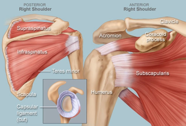
Anterior Shoulder Tendon Anatomy / The Painful Shoulder Part I Clinical Evaluation American Family Physician : An image depicting shoulder anatomy can be seen below.. Tendons are fibrous cords attached to muscles and bone. Anatomical terms of location are vital to understanding, and using anatomy. Atlas of shoulder mri anatomy. Specifically, the four rotator cuff muscles include the following Most common finding is 'military patch' (deltoid) anesthesia.
Robin smithuis and henk jan van der woude. Important to rule out axillary nerve injury. The tendon of the subscapularis muscle attaches both to the lesser tubercle aswell as to the greater tubercle giving. Anterior band of ighl (main restraint). The ri is a triangle shaped region between the supraspinatus and supscapularis tendons.
Shoulder Human Anatomy Image Function Parts And More from img.webmd.com In this episode of eorthopodtv, orthopaedic surgeon randale c. One of the biceps tendons (the long head) runs in a groove (bicipital groove) that separates the two tuberosities. Majority of anterior shoulder dislocations are due to trauma. Latarjet procedure performed more commonly than bristow. 1 enumerate the layers of anterior abdominal wall. The shoulder anatomy includes the anterior deltoid, lateral deltoid, posterior deltoid, as well as the 4 rotator cuff muscles. Dynamic anterior shoulder stabilization with the long head of the biceps tendon: Visit www.handcare.org for more information about conditions, injuries and treatment of the hand, arm, elbow and shoulder.
Irreducible anterior dislocation of the shoulder due to interposition of the long head of bíceps tendón and avulsed part of the labrum, treated arthroscopically;
Subscapularis tendon (open arrow) and anterior labrum (arrowhead) are also shown on this section. Visit www.handcare.org for more information about conditions, injuries and treatment of the hand, arm, elbow and shoulder. Robin smithuis and henk jan van der woude. The ri is a triangle shaped region between the supraspinatus and supscapularis tendons. Normal anatomy, variants and checklist. Provides static restraint with arm in 90° of abduction and external rotation. Anterior graphic of the shoulder. Corey chakarun from shin imaging in california. Related online courses on physioplus. Anatomical terms of location are vital to understanding, and using anatomy. Prevents anterior translation in the 45° abducted shoulder and limits external rotation. Tendons are fibrous cords attached to muscles and bone. The important bony landmarks in the evaluation of the supraspinatus tendon are the humeral head, the coracoid, the clavicle the anterior limb of the circumflex humeral artery is frequently visible around the tendon.
• review pertinent anatomy and pathology associated with common causes of shoulder pain. • pain and/or pop at anterior shoulder but usually not painful after initial event. Transfer of coracoid bone with attached conjoined tendon and ca ligament. Subscapularis tendon (open arrow) and anterior labrum (arrowhead) are also shown on this section. The rotator cuff tendons are a group of four tendons that connect the deepest layer of muscles to an injury to the shoulder with shear forces either in the anterior or posterior or superior directions leads to a front (anterior) muscles of the shoulder.
Shoulder Muscles Anatomy And Functions Kenhub from thumbor.kenhub.com Corey chakarun from shin imaging in california. Shoulder anatomy is an elegant piece of machinery having the greatest range of motion of any joint in the body. Anterior graphic of the shoulder. Muscles of the anterior shoulder. Latarjet procedure performed more commonly than bristow. 1 enumerate the layers of anterior abdominal wall. Pectoral, anterior shoulder, anterior arm. The pectoralis minor muscle is a small.
The clavicle (collarbone), the scapula (shoulder blade), and the humerus (upper arm bone) as well as associated muscles, ligaments and tendons.
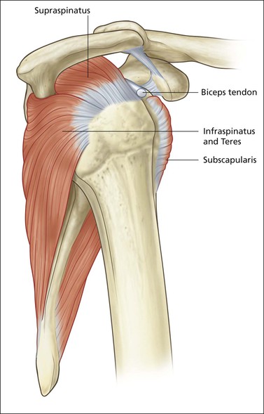
Related online courses on physioplus. Anterior band of ighl (main restraint). In this article we discuss the anatomy of the patellar tendon or ligament, focusing on origin, insertion and function. 1 enumerate the layers of anterior abdominal wall. Sechrest, md narrates an animated tutorial on the basic anatomy of the shoulder. Anterior static shoulder stability is provided by. Infraspinatus and teres minor tendon. Dynamic anterior shoulder stabilization with the long head of the biceps tendon: Subscapularis tendon (open arrow) and anterior labrum (arrowhead) are also shown on this section. 3 what are the vertebral levels of important. Anterior graphic of the shoulder. Normal anatomy, variants and checklist. Shoulder girdle upper limb anterior muscles ligaments tendons posterior views patient positioned rotated prone move figure radiologykey.
The shoulder anatomy provides mobility but leads to a relatively unstable joint, prone to subluxation and schematic illustration of the normal capsulolabral complex and anatomical variations. Shoulder girdle | radiology key. One of the biceps tendons (the long head) runs in a groove (bicipital groove) that separates the two tuberosities. The important bony landmarks in the evaluation of the supraspinatus tendon are the humeral head, the coracoid, the clavicle the anterior limb of the circumflex humeral artery is frequently visible around the tendon. Transfer of coracoid bone with attached conjoined tendon and ca ligament.
Shoulder 1 Supraspinatus Tendon Radiology Key from radiologykey.com The tendon of the subscapularis muscle attaches both to the lesser tubercle aswell as to the greater tubercle giving. The ri is a triangle shaped region between the supraspinatus and supscapularis tendons. Atlas of shoulder mri anatomy. Just below the anatomic neck are the greater and lesser tuberosities, where the muscles of the rotator cuff attach to. Robin smithuis and henk jan van der woude. Pectoral, anterior shoulder, anterior arm. This webpage presents the anatomical structures found on shoulder mri. Corey chakarun from shin imaging in california.
Simple, easy notes for quick revision of important questions.
Irreducible anterior dislocation of the shoulder due to interposition of the long head of bíceps tendón and avulsed part of the labrum, treated arthroscopically; The posterior compartment of the forearm (generally) contains… ___ is caused by a disruption in the extensor tendon. Shoulder girdle | radiology key. Provides static restraint with arm in 90° of abduction and external rotation. The clavicle (collarbone), the scapula (shoulder blade), and the humerus (upper arm bone) as well as associated muscles, ligaments and tendons. • review pertinent anatomy and pathology associated with common causes of shoulder pain. Anterior graphic of the shoulder. Visit www.handcare.org for more information about conditions, injuries and treatment of the hand, arm, elbow and shoulder. The brachial artery lies medial to the biceps tendon. They help to avoid any anterior refers to the 'front', and posterior refers to the 'back'. Most common finding is 'military patch' (deltoid) anesthesia. Learn this topic now at kenhub. The long biceps tendon arises from the supraglenoid tubercle and partly from the superior glenoid labrum (7a).
Glenohumeral joint glenohumeral joint the glenohumeral joint is a multiaxial synovial ball and socket joint and involves articulation between the glenoid fossa of the shoulder tendon anatomy. Shoulder girdle upper limb anterior muscles ligaments tendons posterior views patient positioned rotated prone move figure radiologykey.